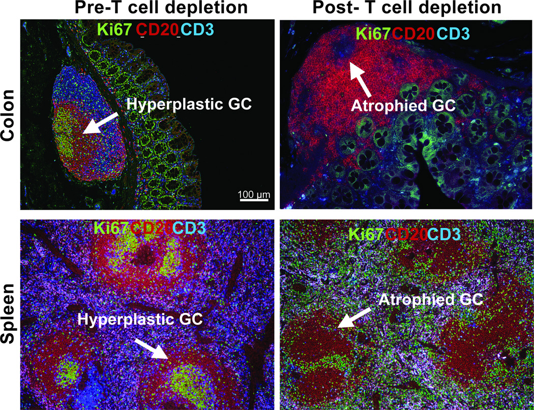
FIGURE 5. Disrupted GC after T cell depletion in spleen and colon.
Representative IHC panels show sections of colon (Upper panel) and Spleen (lower panel) from before and After T cell depletion with large lymphoid aggregation (Peyer’s patch) stained with Ki67, CD20, and CD3 antibodies. (A) Similar to the lymph node, Peyer’s patch and spleen comprises T cell area and B cell follicle with germinal center containing large proliferating B cells. (B) GC in payer’s patch in the descending colon and spleen of rhesus macaques were disrupted after T cell depletion.

