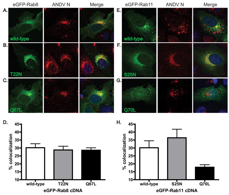
Figure 5. Dominant negative and constitutively active forms of Rab11 have altered levels of colocalization with ANDV N during infection.
ANDV-infected Vero cells were transfected with cDNAs expressing (A) wild-type, (B) T22N mutant, GDP-bound dominant negative, and (C) Q67L mutant, GTP-bound constitutively active, forms of eGFP-Rab8 (green); and (E) wild-type, (F) S25N mutant, GDP-bound dominant negative, and (G) Q70L mutant, GTP-bound constitutively active, forms of eGFP-Rab11 (green). At 18 hpt, cells were fixed and immunostained for ANDV N (red, AlexaFluor 555). Nuclei were counterstained with TO-PRO-3 (blue) and are shown in the merged images only. All images are flattened reconstructions of confocal z-stacks acquired at 63x optical magnification and 2x digital zoom. Levels of colocalization between ANDV N and (D) eGFP-Rab8 and (H) eGFP-Rab11 proteins were quantitated using the Volocity imaging software. Data presented is the mean of three independent experiments and at least 5 cells per cDNA transfection in each experiment.

