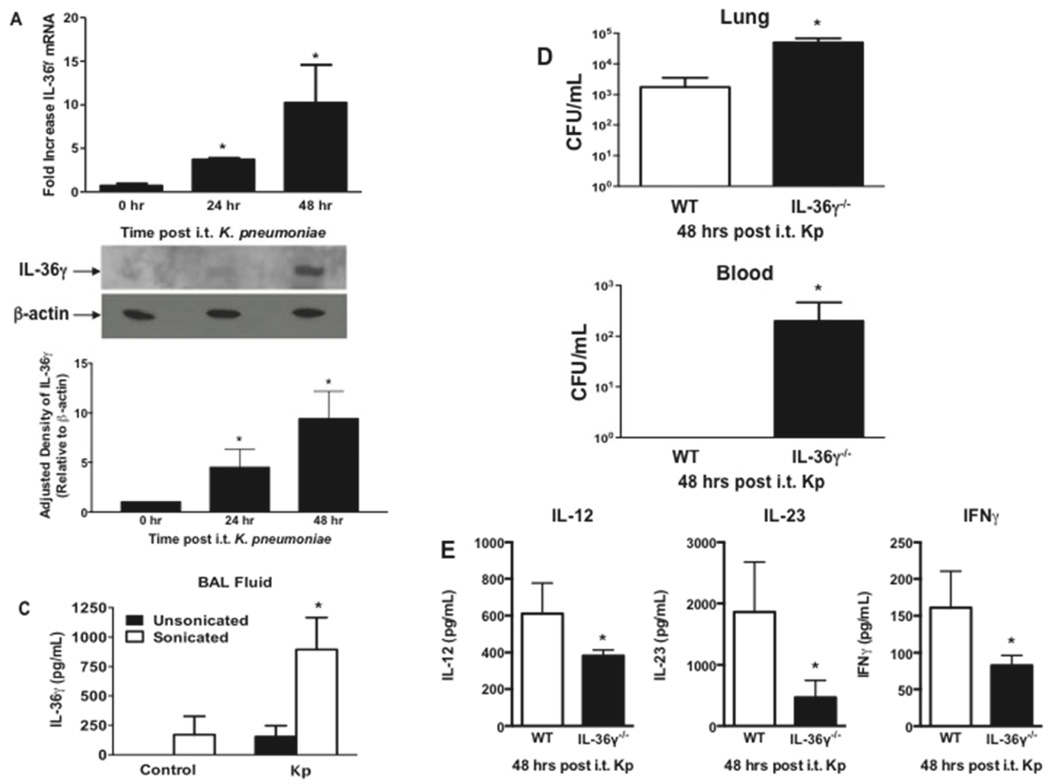
Fig. 9. Effect of IL-36γ on innate immune response to K. pneumoniae.
WT mice were infected with i.t. Kp and lungs were harvested at the specified time points. (A) mRNA was assessed by RT-PCR (*p < 0.05 compared to uninfected controls by one-way ANOVA with Dunnett’s multiple comparisons test, n = 4 mice/group, representative of 3 experiments). (B) IL-36γ protein was detected by Western immunoblotting at 48 hrs after infection. Blot is representative of 3 mice/time point, and of 2 separate experiments. Densitometry represents relative density compared to β-actin from all replicates pooled from both experiments. (*p < 0.05 compared with uninfected mice by one-way ANOVA with Dunnett’s multiple comparisons test). (C) WT mice infected with Kp underwent BAL at 48 hrs after infection. BAL fluid was analyzed for IL-36γ protein by ELISA either with or without sonication. (*p < 0.05 compared to unsonicated control by one-way ANOVA with Dunnett’s multiple comparisons test, n = 4–5 mice/group, representative of 2 experiments) (D) Lung and spleen CFU were assessed by serial dilution at 48 hrs after infection. (*p < 0.05 compared to WT by student’s t test, n = 8 mice/group, combined from 2 experiments) (E) BALF of WT and IL-36γ−/− mice was analyzed for cytokine concentration by ELISA 48 hrs after infection (*p < 0.05 compared to WT by student’s t test, n = 4 mice/group, representative of 2 experiments).

