
Figure 3. Particle adhesion to inflamed mesentery endothelium as a function of combined sLeA and aICAM sites.
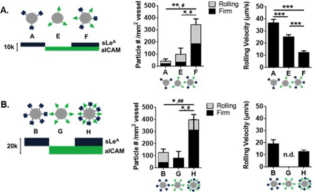
Comparison of dual-targeted particle designs with single ligand densities of (A) 5,000 sites/µm2 and (B) 10,000 sites/µm2Left: Diagram of particle conditions with varied amounts of sLeA and aICAM ligand density. Middle: Quantified adherent density of firmly bound and rolling particles per representative imaging segment, n = 3 mice. Left: Velocity of rolling particles found at mesentery wall, n ≥ 50 particles from n = 3 mice. Statistical analysis of adherent density was performed using one-way ANOVA with Fisher’s LSD test between total adherent particles (*) indicates p<0.05, (**) p<0.01 and two-way ANOVA with Fishers LSD test between groups, (#) indicates p<0.05, (##) p<0.01 between firm groups, rolling groups n.s. Statistical analysis of rolling velocity was performed using one-way ANOVA with Fisher’s LSD test, (***) indicates p<0.001. Error bars represent standard error.