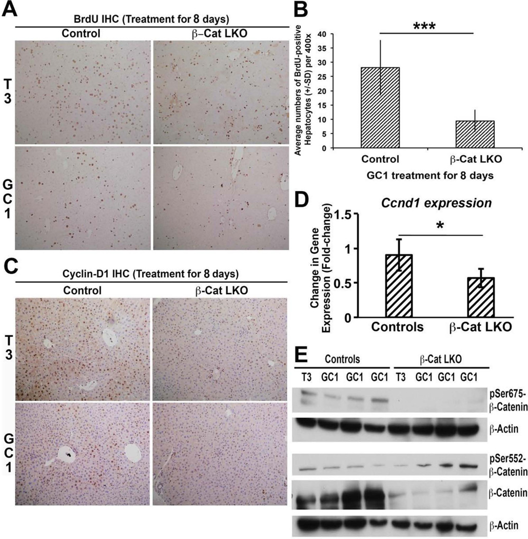
Figure 4.
β-Catenin in hepatocytes is required for GC-1-induced hepatocyte proliferation. (A) Immunohistochemistry for BrdU shows decreased staining in T3 diet-fed and GC-1-injected β-cat-LKO mice as compared to control mice. As shown in previous work, there is increased proliferation in control mice fed T3 diet (100×). (B) Bar graphs showing average number of BrdU-positive hepatocyte nuclei per HPF (400×) in GC-1-injected β-cat-LKO as compared to control mice. A total 10 HPFs per group were counted. A significant difference in hepatocyte proliferation was observed between the two groups (***p < 0.001). (C) Immunohistochemistry for cyclin D1 shows increased staining in the livers of littermate control mice that received T3 or GC-1 as compared to notably less staining in β-cat-LKO mice (100×). (D) Real-time PCR shows significant decrease in mRNA expression of cyclin D1 in GC-1-injected β-catenin-LKO mice as compared to littermate controls (one-tailed t-test, *p < 0.05). (E) Representative Western blots using liver lysates from β-cat-LKO and controls after treatment with T3 or GC-1. As expected, β-catenin and pSer675-β-catenin levels are absent or low. Interestingly, pSer552-β-catenin levels are similar to wild-type or slightly increased.

