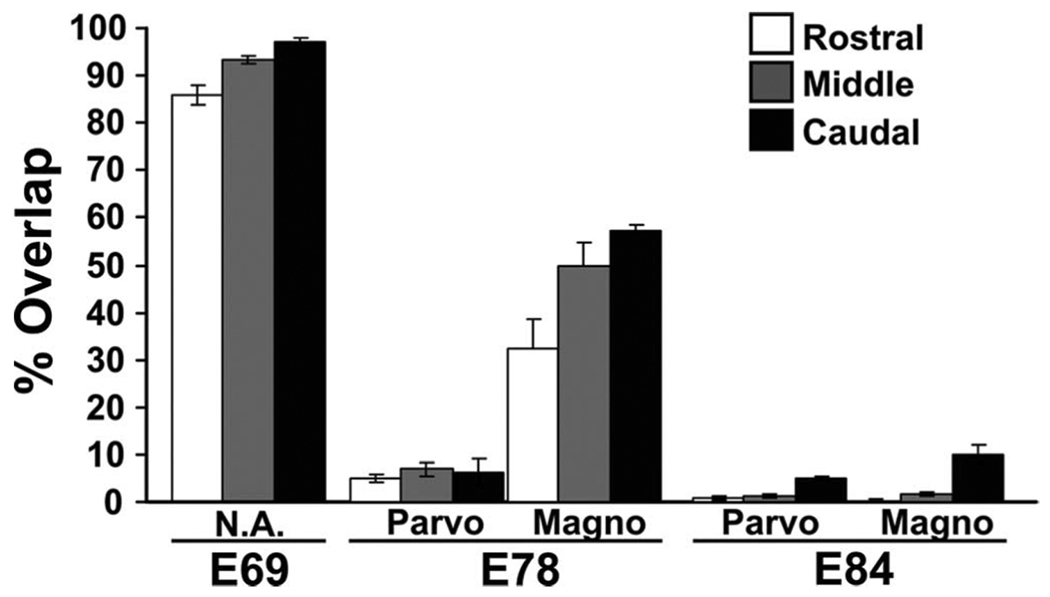
Figure 7.
Quantification of binocular overlap in the developing macaque dLGN. The percentage of the dLGN containing axons from the right and left eyes across the rostral, middle, and caudal dLGN at E69, E78, and E84 is shown. Distinction is made between the degree the parvocellular (Parvo) versus magnocellular (Magno) dLGN on E78 and E84. (TheMvs P regions cannot be reliably distinguished from all portions of the E69 dLGN.) Serial sections through the right and left dLGN were quantified for each age (n=6–10 sections per rostral, 6–10 per middle, and 6–10 per caudal, depending on the size of the dLGN).

