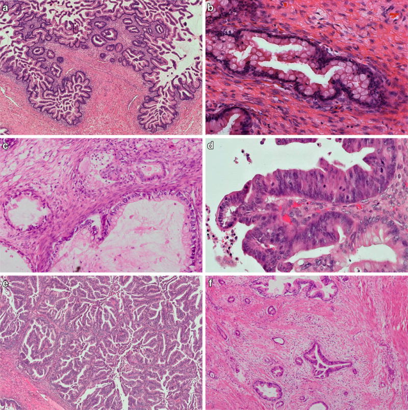
Fig. 2.

MBT showing cystic glandular structures with papillary infoldings, columnar cells with abundant cytoplasmic mucin, admixed with goblet cells of variable degrees of maturation (a) and with basally located nuclei with no considerable nuclear atypia (b). MBT with microinvasion is characterized by small cell groups and glands with cytoplasmic eosinophilia within a normal ovarian stroma without desmoplastic change (c). MBT with intraepithelial carcinoma is characterized by focal high-grade nuclear atypia, commonly associated with more complex epithelial proliferations, next to conventional MBT structures with sharp transition (d). Mucinous carcinoma with expansile (“pushing border”) invasion shows confluent glandular and papillary epithelial proliferations without stromal desmoplasia (e). Mucinous carcinoma with destructive invasion demonstrates haphazardly infiltrating glands and is characterized by desmoplastic tumor stroma (f)
