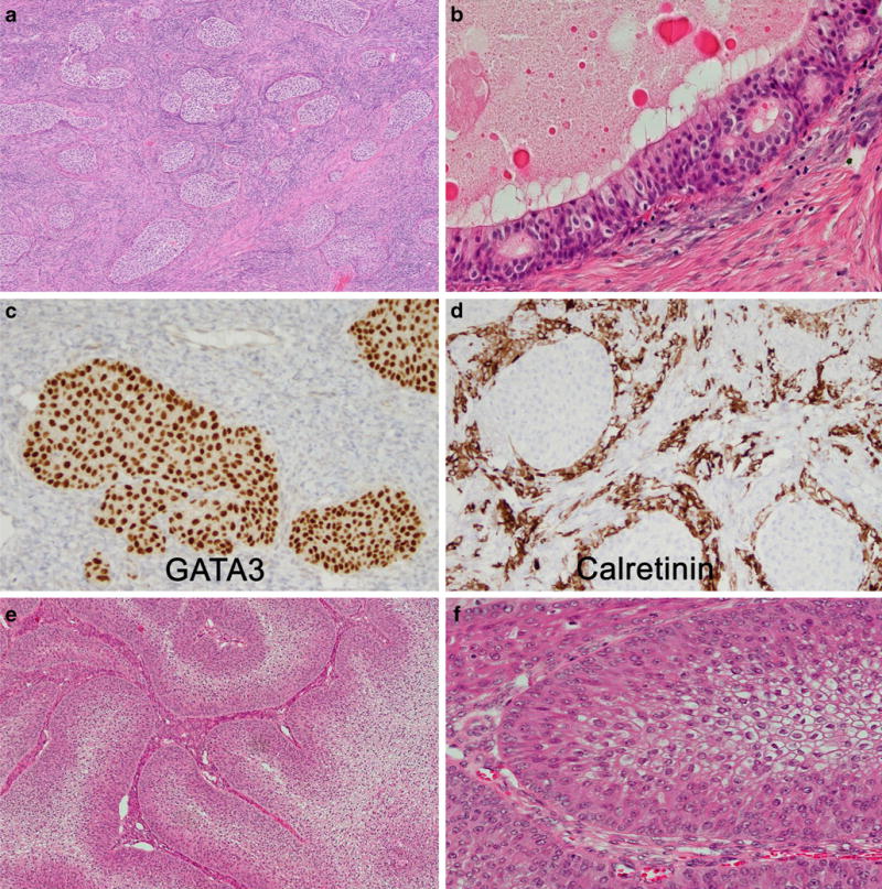
Fig. 4.

Brenner tumor (a) showing epithelial cell nests of variable size, with transitional cell-like morphology, embedded in a fibrous stroma. The epithelium-to-stroma ratio is even. Central cysts are lined by a single layer of columnar mucinous cells. Metaplastic Brenner tumor (b) demonstrates a cystic structure with predominance of mucinous epithelium. GATA3 (c) is diffusely expressed in Brenner tumors, and sometimes many luteinized stromal cells are present highlighted by calretinin stain (d). Brenner BOT are characterized by a significantly increased epithelium-to-stroma ratio (e) but share the same cytological details as benign Brenner tumor (f)
