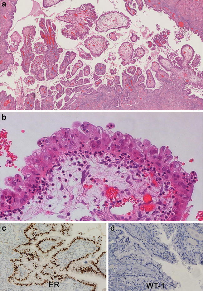
Fig. 5.

SMBT demonstrating a papillary architecture with hierarchical branching (a). The epithelium is columnar with focal multilayering with papillary and pseudopapillary infoldings and variable cytoplasmic mucin content. Stroma and epithelium show infiltration by neutrophils (b). Immunohistochemical expression of ER (and/or PR) (c) and absence of WT1 (d) is typical
