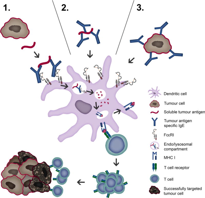
Figure 2. Tumour antigen uptake and presentation by dendritic cells recruits cytotoxic CD8+ T lymphocytes.

Tumour cells display tumour antigens at a high density, facilitating crosslinking of IgE fixed to FcεRI receptors on antigen presenting cells, such as DCs. Tumour antigens may be taken up via three possible routes: 1. soluble tumour antigen binding to receptor-bound IgE; 2. By IgE-opsonized soluble antigen binding to IgE receptors and 3. IgE-opsonized tumour cells binding to IgE receptors. Endocytosis of IgE-antigen complexes leads to digestion in lysosomes and loading of antigenic peptides on MHC I molecules. Cross-presentation via proteasome, loading to MHC I and recognition by CTLs is depicted.
