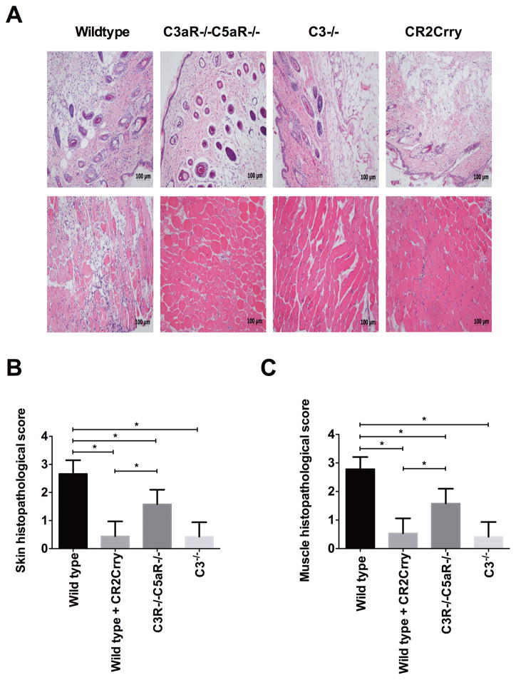
FIGURE 1.
Assessment of IRI damage in vascularized composite allografts from different recipient mice or treated with CR2Crry. Upper panel shows representative H&E stained sections. Lower panel shows histological quantification of injury in grafts. The grafts were isolated at 48h posttransplant. Results are expressed as mean ± SD; n=5–8 for all groups. *P < 0.05

