Figure 4.
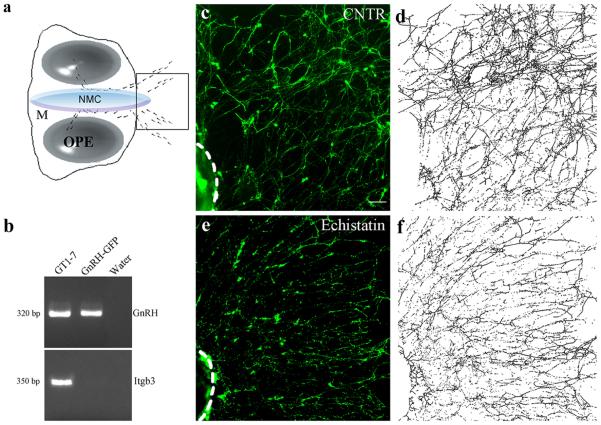
β1-integrin inhibition disrupts GnRH fibers network in vitro. a, Schematic of a nasal explant removed from an E11.5 mouse and maintained in serum-free media for 7 DIV. Ovals represent olfactory pit epithelium (OPE); in center is nasal midline cartilage (NMC) and surrounding mesenchyme (M). GnRH neurons (dots) migrate from OPE and follow olfactory axons to the midline and off the explant into the periphery. Boxed region within schematic is area shown in c–f. b, Representative gel of PCR products for GnRH and β3-integrin (Itgb3) from GnRH neurons isolated from GnRH::GFP E12.5 nasal regions through FACS. Positive (GT1–7 cells) and negative controls (water) were included in the reaction mix. c, e, Explants in experimental groups were maintained in SFM (CNTR) with or without Echistatin (0.1 mM) at 3 DIV for 72 h and fixed at 7 DIV for immunocytochemical processing (GnRH, green). d, f, Representative images binarized and subjected to the “Skeletonize” function of ImageJ software. Scale bars: (in c) c–f, 40 μm.

