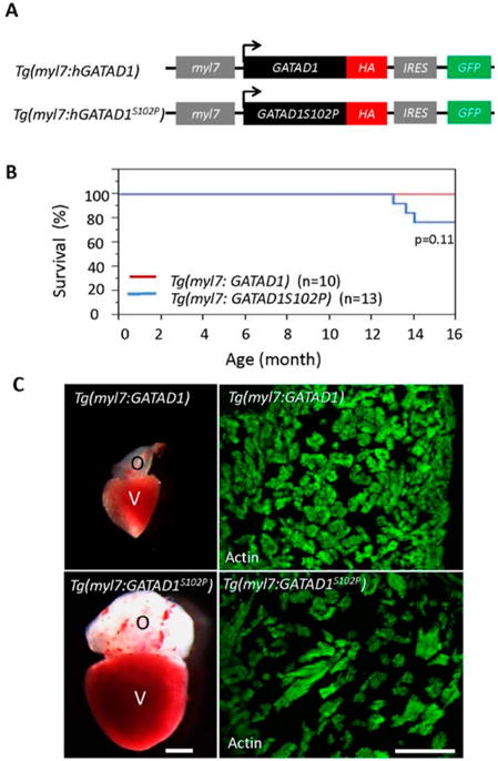
Figure 5.

Generating and phenotyping GATAD1 transgenic fish. (A) Schematic illustration of constructs that were used to generate two transgenic fish lines expressing either human wild type GATAD1 or GATAD1-S102P mutation. The GATAD1 gene is flanked by the myl7 enhancer at its 5′ terminal to drive cardiomyocyte-specific expression, and it has an HA tag and IRES-EGFP at its 3′ terminal to facilitate detection of ectopic gene expression and fish propagation; (B) The transgenic fish expressing mutant GATAD1 started to die at approximately 12 months of age, and all transgenic fish expressing wild type GATAD1 were able to survive to 16 months; (C) Severe cardiac hypertrophy in a Tg(myl7:hGATAD1S102P) fish. Left panel, images of a heart dissected from a Tg(myl7:hGATAD) and a Tg(myl7:hGATAD1S102P) fish at 17 months of age, separately. The heart from this single Tg(myl7:hGATAD1S102P) fish exhibits significantly enlarged ventricle and out flow tract. Right panel, phalloidin staining revealed less dense myofibril and wider myofibril in the heart of this single Tg(myl7:hGATAD1S102P) fish. V, ventricle; O, out flow tract; Scale bar 0.5 mm.
