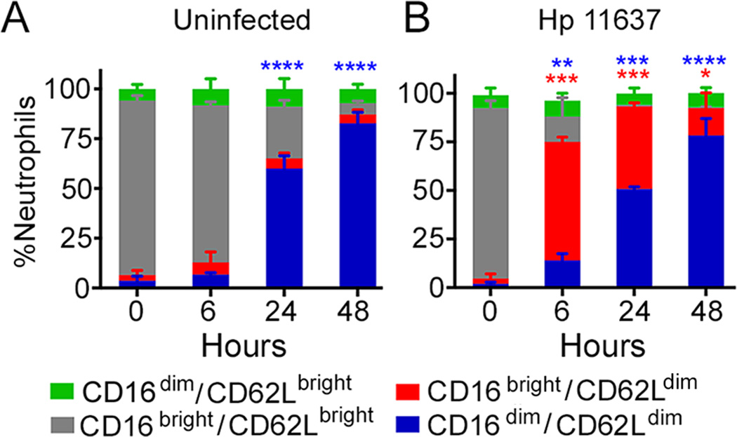
FIGURE 3.
H. pylori-infected neutrophils have high surface CD16 and low surface CD62L. Control, uninfected (A), and H. pylori 11637-infected (B) PMNs were stained for CD16 and CD62L at the noted time points and analyzed by flow cytometry. The percentage of neutrophils in each category is shown as the mean + SEM, n=3. *P<0.05, **P<0.01, ***P<0.001, ****P≤0.0001 vs. 0 h.

