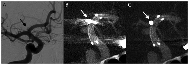
Figure 6.
Illustrative case 2. Immediate DSA (A) maximum intensity projection (MIP) of VasoCT data without MAR (B) and with MAR (C) of stent-assisted coil embolized aneurysm at the right A1 segment. Visibility is significantly affected by streak artifacts caused by the coil mass (arrows) and contralateral clip in VasoCT without MAR. With MAR, stent apposition to vascular wall is fully appreciated.

