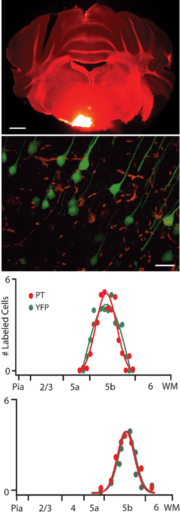
Figure 1. YFPH cells project through the Pyramidal Tract (PT).
Fluorescent microspheres deposited in the Pyramidal Tract (top panel) are retrogradely transported to the somata of YFP-expressing layer 5 pyramidal cells (middle panel). Within the area most densely labeled by microspheres, 42.9 ± 9% of YFP cells (n=92) and 53.6 ± 11% of retrogradely labeled cells (n=87) are double-labeled (n=4 FOV from 2 animals). The laminar distributions in both M1 and S1 of retrogradely labeled and YFP cells overlap, are largely confined to layer 5b, and are not statistically different (bottom panels). Scale Bars indicate 1mm (top) and 25µm (middle).

