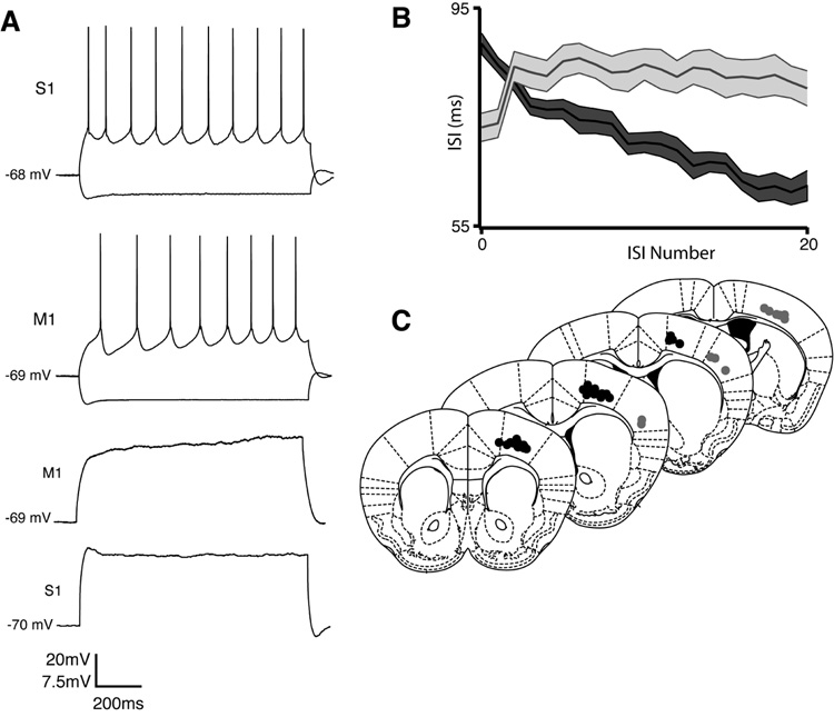
Figure 2. The intrinsic membrane properties and firing types of PT-projecting layer 5 pyramidal cells are regionally distinct.
A. Whereas PT-projecting cells in S1 produce non-adapting trains of action potentials in response to current injection, their counterparts in M1 exhibit a delayed first spike and subsequent spike-frequency acceleration. Subthreshold current injection produces a depolarizing voltage ramp in M1 but not S1. B. Population inter-spike interval (ISI) curves from M1 (dark grey, n=39) and S1 (light grey, n=16). The ratio of the second to last ISI, a measure of spike-frequency adaptation, is statistically different between regions (p < 0.01). Envelopes indicate SEM. C. Somal locations of reconstructed accelerating (dark circles) and non-adapting cells (light circles) with respect to cortical regional boundaries. Accelerating cells are found exclusively in M1 and on the M1/M2 border.

