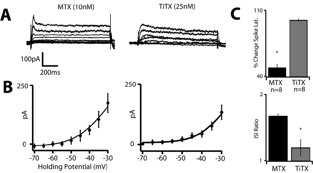
Figure 5. MTX and TiTX-sensitive currents contribute to distinct features of M1 cells' firing type.
A. MTX (10nM) and TiTX (25nM) sensitive currents in M1 PT-projecting cells. Whole-cell current was measured in voltage clamp by stepping to holding potentials between −70 and −30mV from −80mV and the drug-sensitive currents were obtained by subtraction. The MTX-sensitive current includes a transient component that decays rapidly, with no further inactivation, whereas the TiTX-sensitive current decays slowly. B. Population IV curves for the MTX-sensitive current (left, n=9) and TiTX-sensitive current (right, n=10). Error bars indicate SEM. C. MTX but not TiTX significantly (p < 0.01) decreases the latency to first spike (top graph), while TiTX but not MTX significantly (p < 0.01) attenuates spike-frequency acceleration (bottom graph). Error bars indicate SEM.

