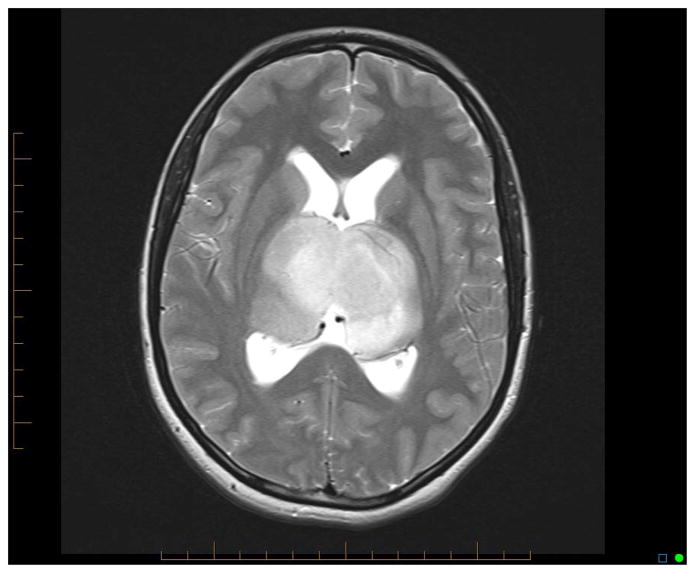
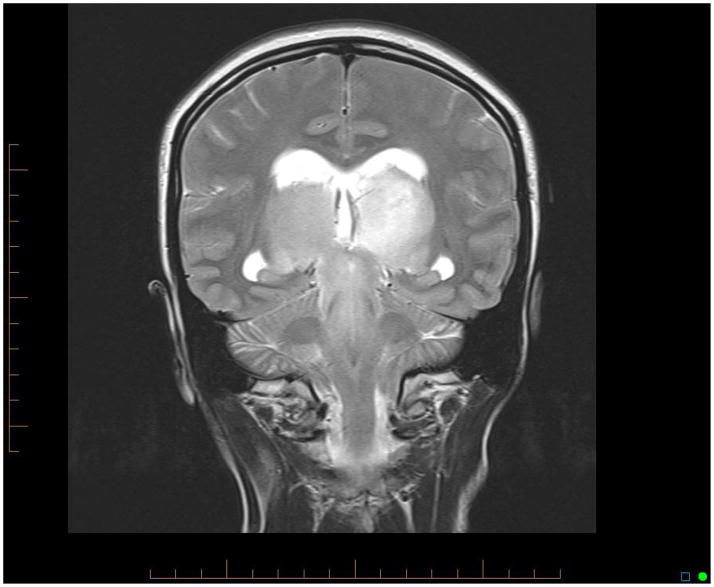
Figure 1.
Axial T2-weighted brain MRI of patient 11 showing symmetrical bithalamic involvement (A). This patient was also found to have hydrocephalus based on symptoms and enlargement of lateral ventricles with transependymal flow in their frontal horns. Coronal T2-weighted brain MRI of same patient showing symmetrical bithalamic and midbrain involvement (B).


