
Figure 2. Structures of viral RNA-directed RNA polymerases.
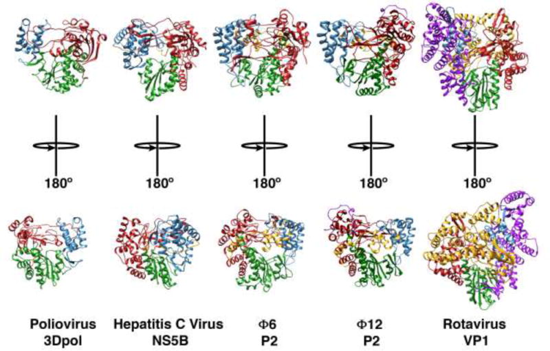
The structures of several viral RdRps are shown in ribbon representation. The structures have been ordered left-to-right with increasing size: poliovirus 3Dpol (PDB: 1RA6, 461 residues, 52.5 kDa) (Thompson and Peersen, 2004), HCV NS5B (PDB: 5CZB, 563 residues, 62.5 kDa) (Pierra Rouviere et al., 2016), Φ6 P2 (PDB: 1HHS, 664 residues, 74.8 kDa) (Butcher et al., 2001); Φ12 P2 (PDB: 4GZK, 659 residues, 75.3 kDa) (Ren et al., 2013) and rotavirus VP1 (PDB: 2R7Q, 1095 residues, 126.1 kDa) (Lu et al., 2008). The fingers, thumb and palm domains are shown in red, blue, and green, respectively. The N- and C-terminal domains, when present, are shown in magenta and yellow, respectively.