
Figure 4. Conserved RdRp sequence motifs.
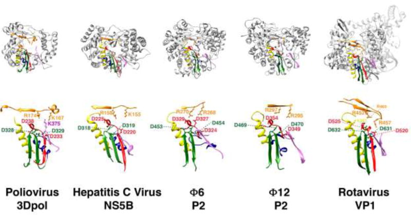
The RdRp sequence motifs A-E (palm) and F (fingers) are mapped onto the structures shown in Figure 2. Motifs A, B, C, D, E and F are shown in red, yellow, green, blue, pink and orange, respectively. A subset of key conserved charged residues is shown in ball-and-stick representation and labeled.