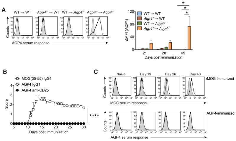
Figure 4.
The naive T-cell and B-cell repertoires of WT mice are purged of AQP4 reactive clones. (A) Criss-cross BM chimeras of WT C57BL/6 and Aqp4−/− mice were immunized with full-length AQP4 protein emulsified in CFA. Sera from different time points after immunization were tested for AQP4-specific antibodies in a cell-based flow cytometric assay as described in Fig. 3. Representative histograms (left) and Mean ΔMFI ± SD (n = 6 per group) for AQP4-specific IgG (right). *p < 0.05 (ANOVA plus Sidak’s post-test). (B) WT C57BL/6 mice were immunized s.c. with full-length AQP4 protein or MOG(35–55) emulsified in CFA as immunization control. On days –5 and –3 prior to immunization, some mice were injected i.p. with control IgG1 or with anti-CD25 antibodies to deplete Treg cells before immunization with full-length AQP4 protein. Mean clinical scores ± SEM (n = 6 per group). ****p < 0.0001 (Mann–Whitney U test for nonparametric values). (C) Aqp4ΔT x Tcra−/− mice were generated by i.p. transfer of the mature T-cell compartment of Aqp4−/− mice into Tcra−/− mice. The mice were immunized with full-length rMOG or AQP4 protein emulsified in CFA. Sera of mice from each group were collected at different time points after immunization and tested for MOG- and AQP4-specific antibodies in a cell-based flow cytometric assay as described. Representative histogram plots illustrating the anti-MOG and anti-AQP4 serum responses in Aqp4ΔT × Tcra−/− mice at different time points (n = 6 per group). Data are representative of two independent experiments (C).

