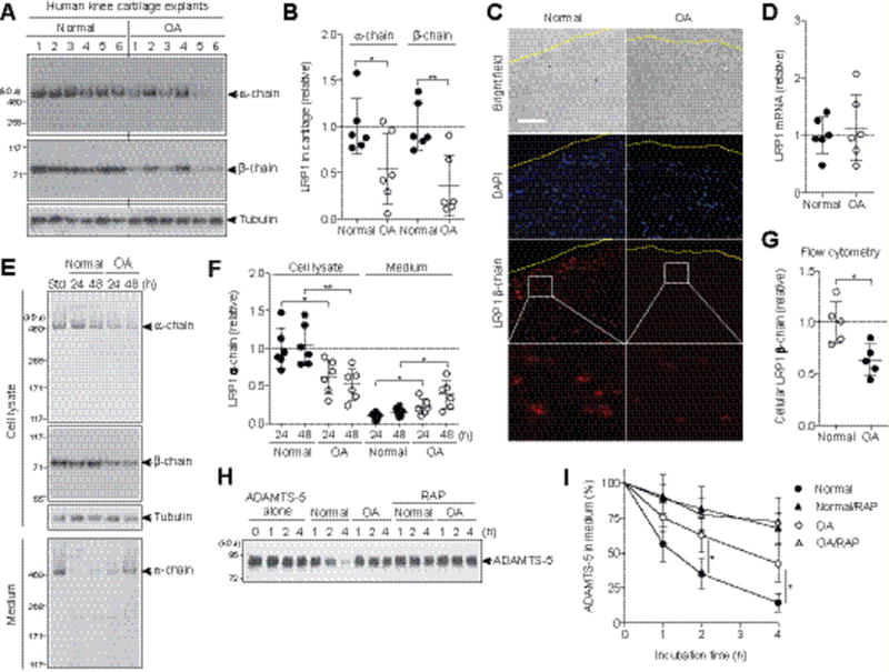
Figure 1. Increased ectodomain shedding of LRP1 and reduced endocytic capacity in human OA cartilage.

(A) The total proteins extracted from the cartilage explants of human knee joints of OA and non-arthritic patients (n=6 each) were subjected to Western blotting with antibodies against α- and β-chains of LRP1. (B) Densitometric analysis of LRP1 in A. Data were normalized against tubulin. (C) Immunoflourescence staining of LRP1 in frozen section of human knee cartilage. Dashed line, articular cartilage surface. Scale bar, 100 μm. (D) Relative LRP1 mRNA levels in cartilage measured by TaqMan qPCR. (E) Representative Western blotting of LRP1 protein in the cell lysates and medium of human normal and OA chondrocytes (n=6 each). std, standard cell lysates. (F) Quantification of LRP1 α-chain detected in E. (G) Flow cytometry analysis of LRP1 β-chain. Each point represents an individual cartilage donor. (H) Representative Western blotting for endocytosis of ADAMTS-5 (10 nM) by human normal and OA chondrocytes (n=3 each) without or with the LRP1 ligand antagonist RAP (500 nM). ADAMTS-5 in the medium was detected by Western blotting using anti-ADAMTS-5 antibody. (I) Quantified data of H. Data are expressed as the mean ± SD. *, p < 0.05, **, p < 0.01, by 2-tailed Student’s t test.
