
Figure 5. The bursicon-expressing neurons of the subesophageal ganglion (BSEG) project throughout the central nervous system.
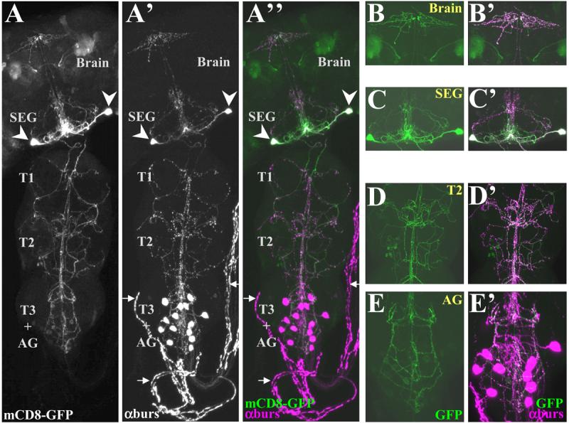
Confocal micrographs of a pharate adult nervous system in which both BSEG neurons have been labeled with mCD8-GFP (green) using the Flp-out system and the Burs-Gal4 driver. The preparation was double-labeled with anti-burs antibody (magenta) to reveal the distribution of bursicon.
(A-A″) mCD8-GFP labeling (A) and anti-burs immunostaining (A′) overlap extensively in the CNS as seen in the merged image (A″), in which. Arrows indicate the abdominal nerves, which lack overlapping GFP (green) and anti-burs (magenta) signals. Arrowheads, somata of the BSEG.
(B-E′) Higher magnification images of BSEG processes in selected regions of the nervous system shown in A. (B-E) mCD8-GFP labeling (green), and (B′-E′) overlappping anti-burs immunostaining in (B,B), brain, (C, C′), subesophageal ganglion, (D,D′), 2nd thoracic ganglion, and (E,E′) abdominal ganglion. Images in A-A″ are volume-rendered z-stacks acquired with a 20X objective representing the entire nervous system. B-E represent limited image planes through the nervous system acquired with a 60X objective.