
Figure 8. Cell death of the BAG is delayed in rk4 mutants.
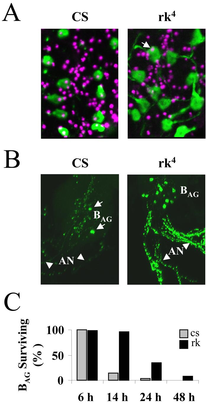
(A) Apoptosis is initiated within six hours of eclosion in the BAG of Canton S control animals (CS, left) but not in those of rickets4 mutants (rk4, right). Each image is a composite of five merged confocal z-sections showing most of the BAG (green) from a representative animal, visualized by anti-burs immunostaining. TUNEL labeling (magenta), which indicates DNA fragmentation, can be seen in the nuclei of all 11 BAG from the control animal (left), but only one of 12 BAG nuclei (arrow) is labeled in the nervous system preparation from a rk4 animal (right). TUNEL-labeled nuclei of many neurons other than the BAG are also visible, consistent with a broader pattern of post-eclosion apoptosis (see Results).
(B) Volume rendered confocal micrographs of the abdominal ganglion and abdominal nerves (AN, arrowheads) from Canton S control (CS, left) and rickets4 (rk4, right) animals immunostained with anti-burs antibody. All BAG are present in the abdominal ganglion of the rk4 mutant, while only two survive in the abdominal ganglion of the control animal (arrows). Substantial bursicon immunoreactivity is associated with the processes of surviving BAG in the abdominal nerves of the rk4 mutants.
(C) Bar graph showing the percentage of BAG neurons still present in animals at the indicated times after eclosion. CS, Canton S control animals; rk4, rickets mutants. Cell counts were averaged from at least six preparations for each genotype and time point.