Figure 7.


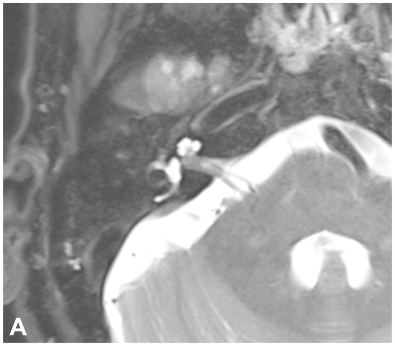
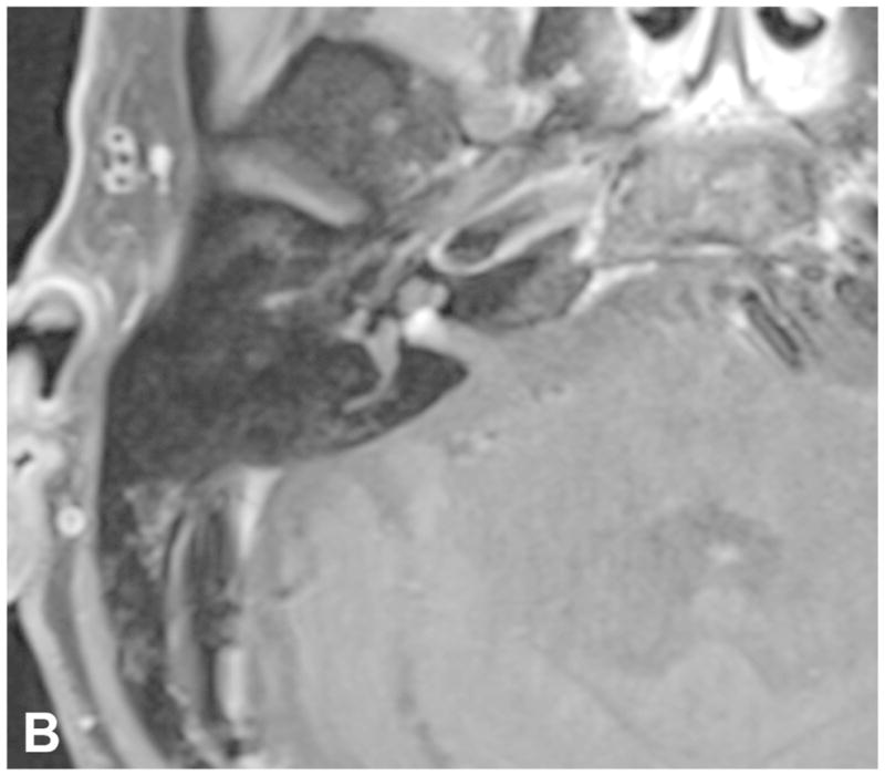
Pre-contrast axial T2-WI (A) and post-contrast axial T1-WI (B) demonstrate a small intracanalicular VS with lateral extension into the IAC fundus as well as the modiolus, which is associated with a decreased rate of hearing preservation.


Pre-contrast axial T2-WI (A) and post-contrast axial T1-WI (B) demonstrate a small intracanalicular VS with lateral extension into the IAC fundus as well as the modiolus, which is associated with a decreased rate of hearing preservation.