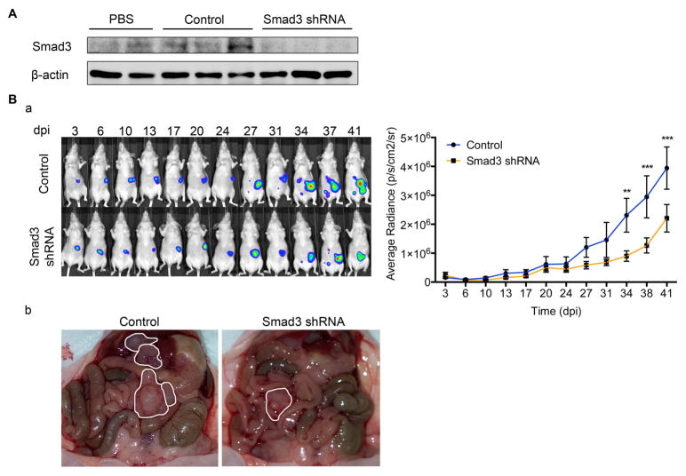
Figure 6. Lentiviral knockdown of Smad3 reduces tumour progression in the peritoneum.
A. Western blot shows the expression level of Smad3 in parietal peritoneum lysates of mice injected in a preliminary assay with PBS (n=2), control lentiviral particles (n=3) or Smad3 shRNA-producing lentiviral particles (n=3). Expression of β-actin was employed as a loading control. B. (a) Representative images of SKOV3-D3 cells bioluminescence in mice pre-conditioned with lentiviral particles producing Smad3 shRNA or control. Quantification of bioluminescence showed that tumour growth was significantly reduced in mice pre-conditioned with lentiviral particles producing Smad3 shRNA (n=9) compared to controls (n=9). Mice of both groups were monitored for 41 days. Graph represents mean average radiance (expressed as photons/s/cm2/sr) of SKOV3-luc-D3 cells ± SEM. Symbols represent the statistical differences over time between both groups (**P ≤ 0.01; ***P ≤ 0.001, P ≤ 0.0001). dpi: days post inoculation. (b) Representative images show a decrease in number of metastases in Smad3 knockdown mice compared to control group. Tumours are outlined in white.

