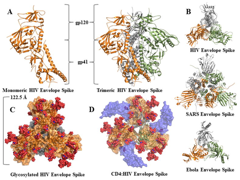
Figure 2. Viral Envelope Protein Complexes.
A) X-ray crystal structures of trimeric Env are rendered in ribbon with each monomer of gp41/120 shown in orange, grey, or green. B) The HIV (4TVP), SARS (5I08), and Ebola (5JQB) Env proteins are rendered in the same fashion, but viewed orthogonally along the three-fold symmetry axis to highlight structural conservation between viruses. C) Glycoproteins were rendered with the glycans as red spheres (4TVP). D) CD4:gp120 were modelled on the Env trimer to demonstrate what the recognition complex would look like (5CAY and 4TVP).

