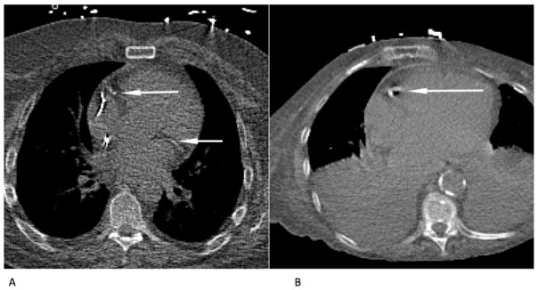
Figure 2.
Examples of calcifications correctly detected by the automatic algorithm in CTAC scans. (a) CAC lesions in the RCA and LCX in CTAC at rest in a scan with metal implants. (b) CAC in the RCA strongly affected by cardiac motion in CTAC scan at stress showing severe abnormalities in the lungs.

