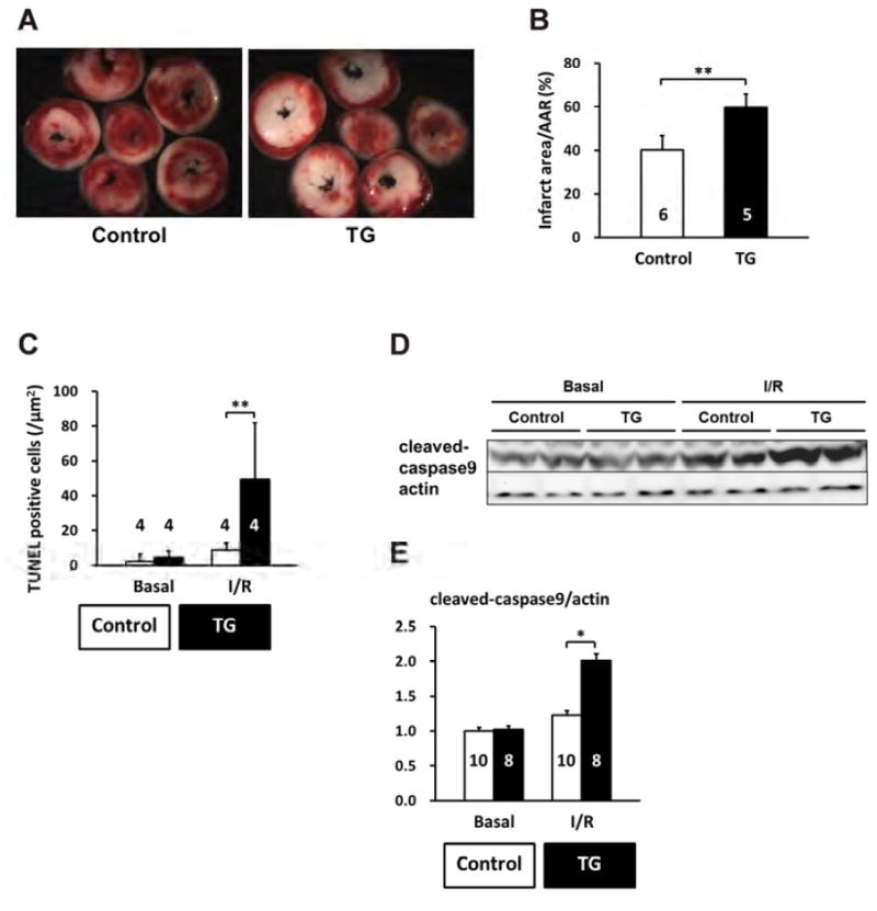
Figure 10. Induced PP2Ce expression promotes cardiomyocyte death in response to ischemia/reperfusion injury.

A. Representative TTC staining cross-section images from one PP2Ce transgenic (TG) and one control (control) heart following 15 minutes of ischemia and 120 minutes of reperfusion (I/R). B. Infarct sizes of control (n=5) and PP2Ce transgenic (TG, n=6) hearts as measured from A. **: p<0.01 between control and PP2Ce TG groups. C. Apoptotic index measured by TUNEL staining in control (n=4) and PP2Ce TG (n=4) hearts. **: p<0.01 between corresponding control and PP2Ce TG groups. D. Representative immunoblot for level of activated caspase9 in control (n=10) and PP2Ce transgenic (TG, n=8) hearts following the same I/R protocol. E. Quantification of level of cleaved caspase9 signal from immunoblots. *: p<0.05 between control and PP2Ce TG groups.
