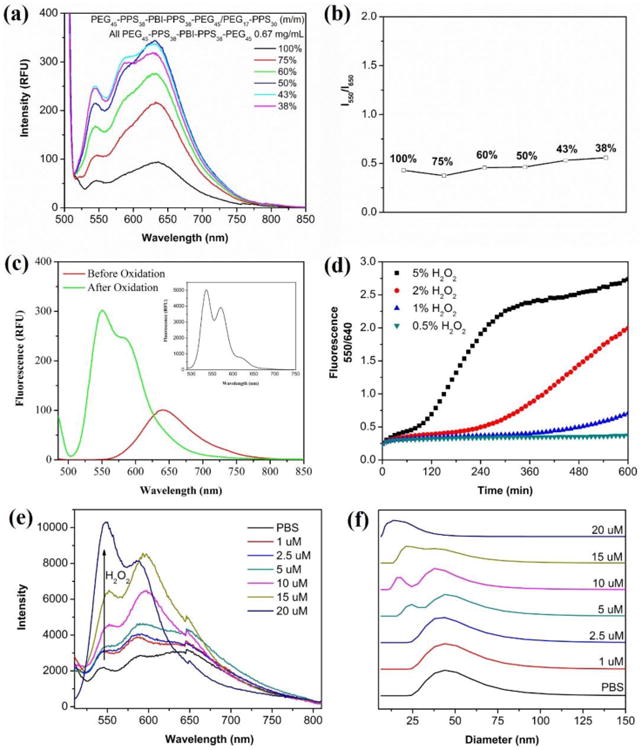
Figure 3.

Characterization of PEG17-PPS28-PBI-PPS28-PEG17 polymersome spectral properties. (a) Red fluorescence emission and (b) red / green emission intensity ratio of polymersomes composed of different ratios of PEG17-PPS28-PBI-PPS28-PEG17 and PEG17-PPS28. The percentage of tetrablock in each tested formulation is shown in the legend. (c) Fluorescence spectra of polymersomes in PBS (2 mg/mL) before and after oxidation by H2O2. The emission in DCM (2 mg/mL) is inserted into the top right corner of the image. (d) Plots of fluorescence intensity ratio at 550 nm and 640 nm as a function of the incubation time at various H2O2 concentrations at 37 °C. RFU = relative fluorescence unit, λex = 485 nm. (e) Emission spectra and (f) vesicle-to-micelle transition of 100% PEG17-PPS28-PBI-PPS28-PEG17 polymersomes after reaction with physiological concentrations of H2O2. Micelle formation was monitored by DLS.
