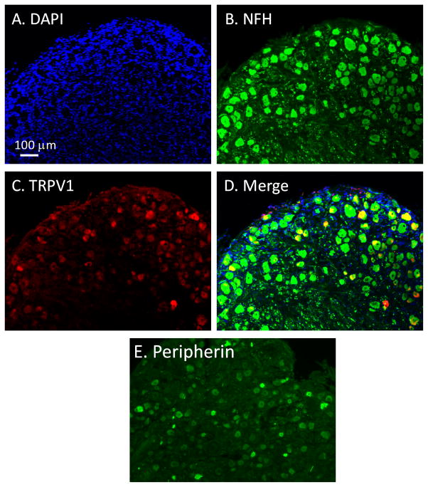
Figure 1. Histochemical characterization of human DRG neurons.
(A) DAPI nuclear staining in an intact hDRG section. (B, C) Double-staining of neurofilament (NF200 or NFH, green, B) and TRPV1 (red, C) in the section. (D) Merged image of DAPI, NFH, and TRPV1 staining. (E) Immunostaining of peripherin in an hDRG section. Scale bar, 100 μm for A-E.

