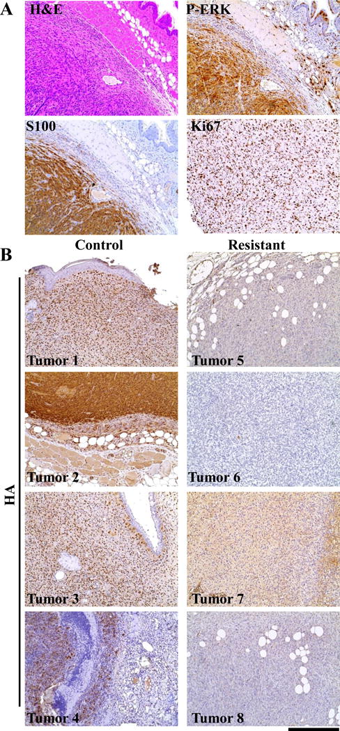
Figure 6. Melanoma histology.

(A) The immunoprofile of representative MEKGF melanoma. MEK melanomas were highly vascular and consisted primarily of short spindle cells exhibiting high-grade nuclear features and prominent nucleoli. They were seen to invade into subcutaneous fat, muscle, and cartilage. IHC for S100 demonstrated the melanocytic origin of the tumors. IHC for P-ERK demonstrated canonical MAPK pathway activation. Assessment of cellular proliferation was performed on slides with uniform tumor cellularity using IHC for the cellular proliferation marker Ki67, demonstrated that all of the tumors were highly proliferative. IHC sections were counterstained with hematoxylin. A Hematoxylin and eosin (H&E) stained tumor section provided for comparison. (B) In situ assessment of MEK expression in Dox resistant tumors. Resistant tumor sections and controls were assessed by IHC for expression of the HA epitope tag on virally delivered MEKGF. All control tumors showed a mixture of nuclear or cytoplasmic HA expression that which not detected in resistant tumors demonstrating continued TET regulated suppression of MEKGF expression with Dox. IHC sections were counterstained with hematoxylin. The scale bar represents 200 μm.
