
Figure 1. CDDP-resistant cells express high levels of AURKA and p-eIF4E (S209) proteins.
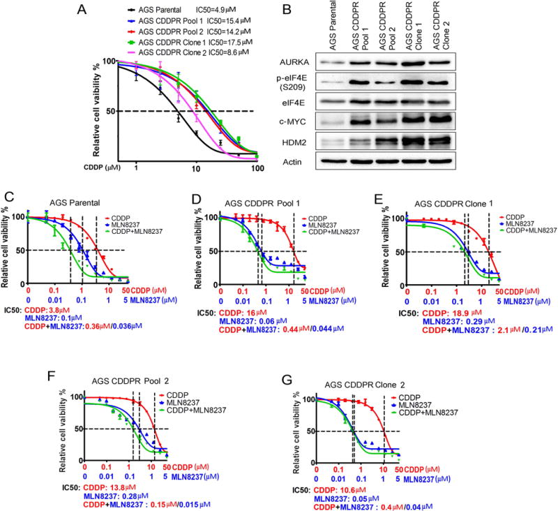
(A) AGS Parental, AGS CDDP-resistant (CDDPR) Pool 1, AGS CDDPR Pool 2, AGS CDDPR Clone 1, and AGS CDDPR Clone 2 cells in 96-well plates were treated with CDDP following a 8-point 2-fold serial dilution, starting at 100 μM concentration. Cell viability was measured using CellTiter-Glo assay. Dose-response was fitted using a three-parameter non-linear regression method. (B) Cell lysates from AGS Parental and CDDP resistant cell lines were subjected to Western blot analysis of the indicated proteins. Levels of AURKA, p-eIF4E (S209), c-MYC, and HDM2 proteins were substantially higher in AGS CDDP-resistant cells than AGS Parental cells. AGS Parental (C) AGS CDDPR Pool 1 (D) and AGS CDDPR Clone 1 (E) AGS CDDPR Pool 2 (F) and AGS CDDPR Clone 2 (G) cells in 96-well plates were treated with MLN8237 alone, CDDP alone, or CDDP + MLN8237 at a fixed ratio (10:1) following a 12-point 2-fold serial dilution. Cell viability was measured and dose response curves were generated as in (A). The mean value of IC50 of each drug treatment was generated from at least three independent experiments.