
Figure 1.
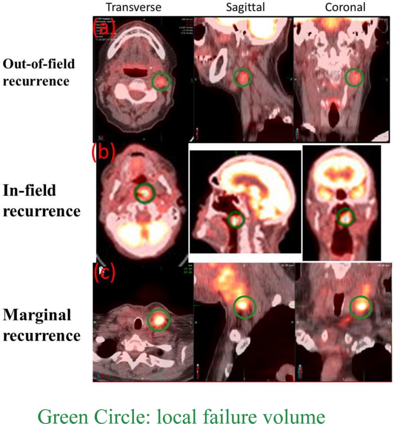
PET/CT scans showing out-of-field (top panel), in-field (middle panel), and marginal local failure (bottom panel) in the transverse (left column), sagittal (middle column), and coronal (right column) plane. The dark green circles indicate the locations of local failure.