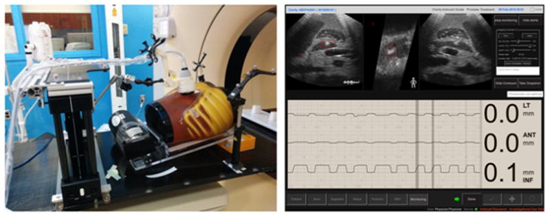
Fig. 11.

The ultrasound phantom, motion platform and arm-bridge system setup in the treatment room (left) and the Clarity real-time monitoring of the ultrasound phantom motion (right). The monitoring module shows the 3D motion with time in left-right, anterior-posterior and superior-inferior directions and real-time ultrasound image views at the top row.
