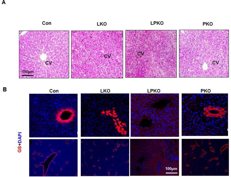
Figure 5. Effect of LKB1 and PTEN loss on liver zonation.
A. H&E staining of liver sections from 3 week old mice shows lack of structure in LPKO livers. B. Staining with glutamine synthetase (GS, red) shows that central venous cells with GS staining are not formed in the LPKO livers. The GS positive cells are also scattered in the LKO livers instead of forming the central vein. CV, central vein. Scale bar, 100µm.

