
Figure 2. Histology and NF-κB expression.
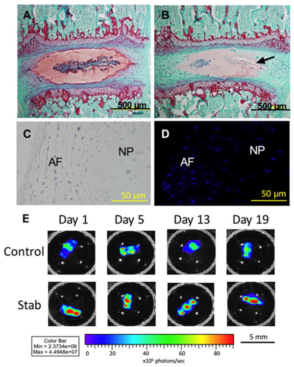
(A) After 21 days in culture, Safranin O staining (red) shows that proteoglycan content is maintained in both the AF and NP in the Control samples. (B) The Stab samples (puncture site indicated by arrow) showed decreased proteoglycan content in both the AF and NP. (C) Tetrazolium blue staining (blue) shows co-localization. (D) DAPI staining shows that cells are metabolically active and viable after 21 days in organ culture. (E) NF-κB expression in both the Control and Stab samples also indicate that the IVD is viable and responsive to its environment. The Stab samples have increased NF-κB expression at the 1, 5, 13, and 19-day time points. Lumbar IVDs are shown here.