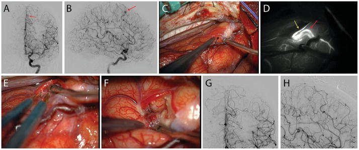
Figure 4.
Case illustration. This 49-year-old lady was evaluated for chronic headaches and a history of recurrent nose bleeds and multiple cutaneous telangiectasias. She had a family history of bleeding lesions in her first and second degree relatives. Antero-posterior (A) and lateral (B) cerebral angiograms with left internal carotid artery injection showed a small left medial frontal arteriovenous malformation (Spetzler-Martin grade 1, Lawton-Young grade 3, Supplemented Spetzler-Martin grade 4) (red arrows). The lesion was exposed through a left interhemispheric approach (C, black arrow). Intra-operative indocyanine green video angiography showed the feeder artery (yellow arrow) and the single arterialized draining vein (red arrow) leading from the nidus (D). The feeding artery was skeletonized, coagulating the small branches supplying the nidus and preserving distal flow in the parent artery. (E) The nidus was circumferentially dissected and removed. Post-operative antero-posterior (G) and lateral (H) angiograms confirmed complete resection of the lesion. The patient was neurologically intact postoperatively.

