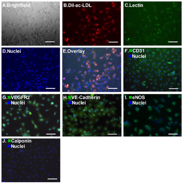
Figure 2. Endothelial Cell Phenotypes.
The attached cells display spindle-shaped and cobblestone-like appearances (A, bar = 100 μm), double-positive fluorescence of Dil-ac-LDL and Ulex-Lectin (B–E, bar = 100 μm). Meanwhile, platelet endothelial cell adhesion molecule 1 (CD31, PECAM-1), VEGFR2, VE-Cadherin, eNOS were positive in >95% of the cells after the second passage (F–I, bar = 100 μm) Most of the cells were negative for Calponin staining, which is a smooth muscle cell marker (J, bar = 100 μm).

