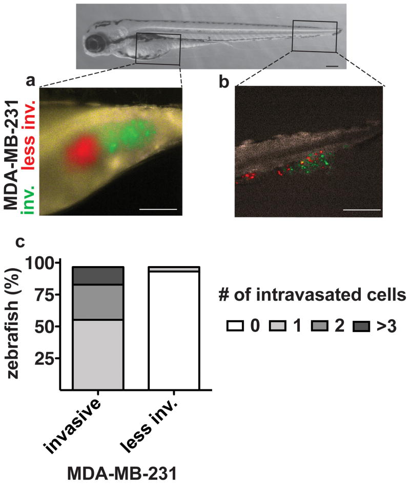
Figure 4. Intravasation of MDA-MB-231 cell populations 2 days after yolk sac injection.
(a) Yolk sac with green (invasive) and red (less invasive) labeled MDA-MB-231 cells. (b) The caudal region with MDA-MB-231 cells that invaded the vasculature to reach the tail region. (c) The number of zebrafish embryos with intravasated MDA-MB-231 cells in the caudal region 2 days after injection26. Scale bars, 250 μm.

