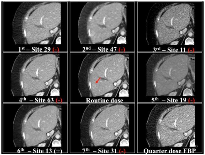
Figure 9.
Demonstration of a true positive image (white +) and false negative images (red −) for a hyperattenuating metastasis shown with the red arrow relative to the commercial filtered backprojection (FBP) and quarter dose FBP images. In this example, readers missed the lesion for all sites except one (site 13).

