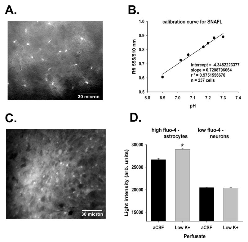
Figure 1.
Expression of EGFP in astrocytes in brain slices prepared from transgenic mice is shown in panel A (scale bar = 30 micron). In panel B, the calibration curve for SNAFL is shown. Cells were exposed to solutions containing nigericin at known pH values ranging from 6.9-7.3 and the ratio of fluorescence at two excitation wave lengths (550 and 510 nm) was determined. The calibration data were fit using a linear regression equation because we used only a small pH range close to the pKa of SNAFL. Fluo-4 loading is shown in panel C (scale bar = 30 micron); the cells that stained more intensely for Fluo-4 were astrocytes. Fluo-4 is a Ca++ sensitive dye, and when the population of Fluo-4 loaded cells was divided into two groups on the bases of the intensity of Fluo-4 fluorescence in aCSF (panel D), only those cells with more intense uptake demonstrated increased intracellular Ca++ levels after exposure to low potassium solutions (Dallwig and Deitmer, 2002). “*” indicates P < 0.05.

