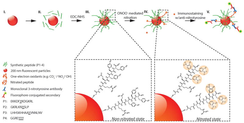
Figure 1.
Schematic representation of peroxynitrite-induced nitration. (I) The fluorescent particle, (II) conjugation of peptides with EDC/NHS crosslinker (P1–P4), (III) non-nitrated peptides conjugated to surface of fluorescent particles, (IV) nitration of tyrosine through peroxynitrite-mediated pathway, (V) immunostaining of nitrated peptides with anti-nitrotyrosine IgGs and fluorescent secondary IgGs. (Steps I–III): Carboxyl-functionalized red fluorescent particles (≈200 nm in size) are coated with tyrosine-containing peptides (P1–P4, green strands). Step IV: Peroxynitrite-mediated nitration of tyrosine residues resulting in the formation of 3-nitrotyrosine. Step V: Immunostaining of nitrated peptides with monoclonal anti-nitrotyrosine IgGs (MAB5404; Millipore) and fluorophore-conjugated secondary IgGs.

