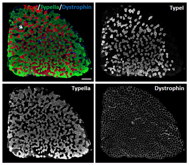
Figure 3. Immunostaining of myofibers in SOL muscles of 2-month old male mice.
Myh2, Myh7 and Dystrophin primary antibodies were used to stain Type IIa (Green), Type I (Red) myofibers and sarcolemma (Blue), respectively. Unlabeled myofibers (Black) are presumed to be Type IIx myofibers. One Type IIx myofiber is indicated by the arrow. Scale bar = 200 μm.

