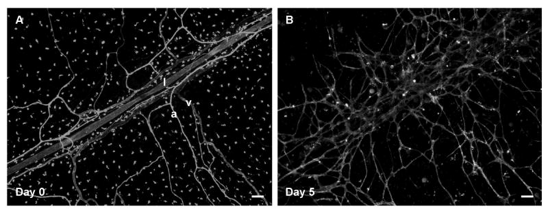
Figure 6. Time-lapse images demonstrate the ability to observe lymphatic and blood vessel patterning.
Lymphatic (l) vessels can be distinguished from arterioles (a) and venules (v) based on labeling morphology on day 0 (A). On day 5 post-stimulation with 10% serum, lymphatic morphology is lost and vessels appear to have integrated with the nearby angiogenic blood vessels. Scale bars = 100 μm.

