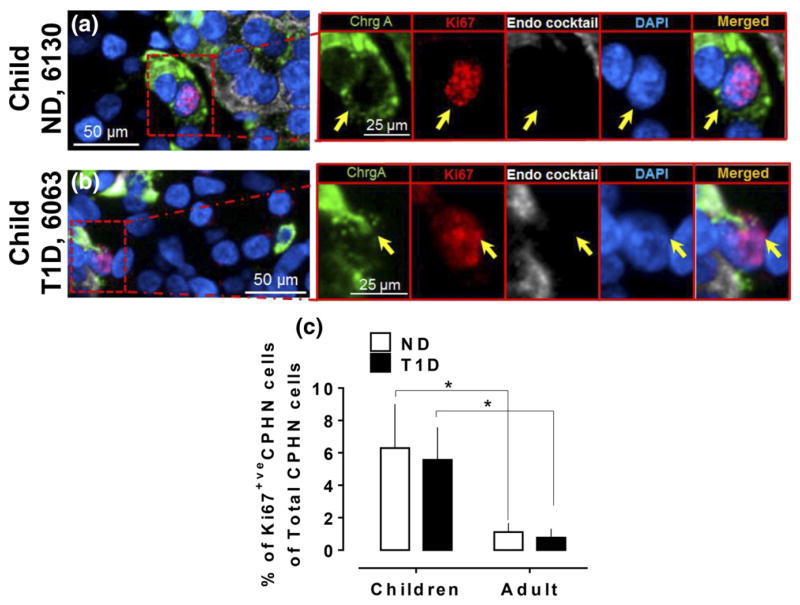
Figure 2.
The frequency of replication of CPHN cells in the pancreas is comparable in children with and without T1D and more frequent than in adults. Representative images of CPHN and Ki67 staining in the pancreas in (a) a child ND donor and (b) a child T1D donor. Individual layers stained for ChrgA (green), endocrine cocktail (insulin, glucagon, somatostatin, pancreatic polypeptide, and ghrelin: white), Ki67 (red), and DAPI (blue) are shown along with the merged image. Yellow arrows indicate CPHN cells. (c) Comparison of Ki67-positive CPHN cells between children and adults (in both T1D and ND) in all compartments of pancreas. Scale bars: 50 μm for lower-power images and 25 μm for insets.

