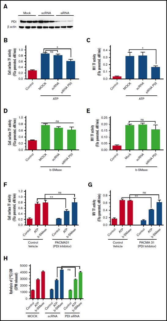
Figure 7. Effect of PDI inhibition on ATP- and b-SMase-induced TF decryption, TF+ MVs release, and SM hydrolysis.
(A) Cell lysates of MDMs transfected with mock, scRNA, or siRNA specific for PDI were probed for PDI expression levels by western blot analysis. (B–C) MDMs transfected with mock, scRNA, or PDI siRNA were treated with a control vehicle or BzATP (200 µM) for 15 minutes. TF activity on the cell surface (B) or MVs (C) was measured in FX activation assay. (D–E) MDMs transfected with mock, scRNA, or PDI siRNA were treated with a control vehicle or b-SMase (1 U/mL) for 1 hour. Following the treatment, cell surface TF activity (D) or TF activity in MVs (E) was measured in FX activation assay. (F–G) MDMs were first treated with a control vehicle or PDI inhibitor PACMA31 (10 µM) for 1 hour, and then treated with control vehicle, BzATP (200 µM, 15 minutes), or b-SMase (1 U/mL, 1 hour). Cell surface TF activity (F) or MVs TF activity (G) was measured in FX activation assay. (H) MDMs transfected with mock, scRNA, or PDI siRNA were metabolically labeled with [methyl-14C]-choline chloride (0.2 µCi/mL) for 48 hours. Labeled MDMs were treated with a control vehicle, BzATP (200 µM, for 15 minutes), or b-SMase (1 U/mL, for 1 hour). Cell supernatants were collected and centrifuged to remove cell debris and MVs before they were counted for the radioactivity to determine the release of [14C]-phosphorylcholine as an index for SM hydrolysis.

