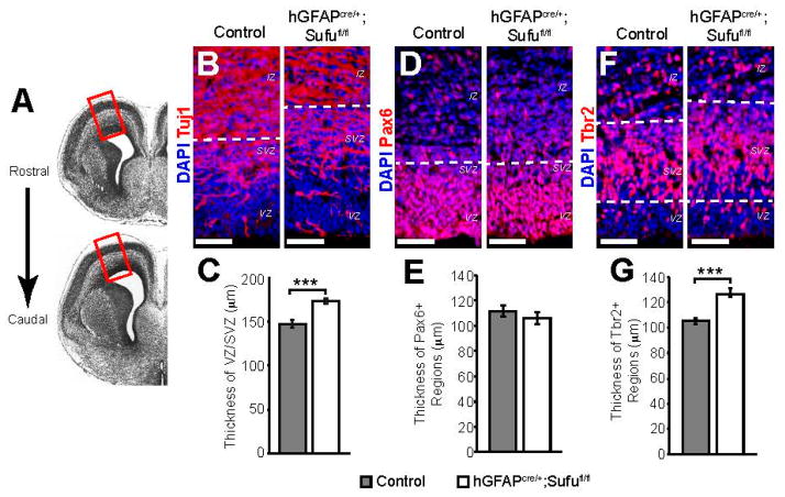
Figure 1.
Expansion of the progenitor zones in E16.5 hGFAPcre/+;Sufufl/fl mice. (A) The experiments presented here analyzed the dorsolateral region of the neocortex (boxed region), obtained from the rostral forebrain spanning between these representative images; (B,C) Targeted deletion of Suppressor of Fused (Sufu) in cortical progenitors at E13.5 (hGFAPcre/+;Sufufl/fl) led to the expansion of the VZ/SVZ by E16.5 as defined by immunofluorescence staining. Regions marked by DAPI-dense (blue) cells and reduced Tuj1 (red) immunostaining along the lateral ventricles showed expansion of the hGFAPcre/+;Sufufl/fl VZ/SVZ compared to controls (B); The thickness of the VZ/SVZ was determined as the distance between the ventricular lining along the lateral ventricles and the IZ, which was identified by densely packed DAPI+ cells and minimal Tuj1 staining within the dorsolateral neocortex. These measurements confirmed that the hGFAPcre/+;Sufufl/fl VZ/SVZ was significantly thicker than those of control littermates (C); (D,E) Immunofluorescence staining against the RG cell marker, Pax6 (red), showed no visible differences between the E16.5 control and hGFAPcre/+;Sufufl/fl Pax6+ regions as defined by the dashed lines (D); This observation was verified when the thickness of Pax6+ regions was measured (E); (F,G) Immunofluorescence staining against the intermediate progenitor (IP) cell marker, Tbr2 (red), showed a visible expansion of Tbr2+ regions (between dashed lines) in the E16.5 hGFAPcre/+;Sufufl/fl VZ/SVZ when compared to controls (F); Quantification of the thickness of Tbr2+ regions verified these observations (G); *** p-value < 0.01. Scale bars = 50 μm; VZ, ventricular zone; SVZ, subventricular zone; IZ, intermediate zone; DAPI, 4′,6-diamidino-2-phenylindole.

