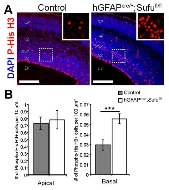
Figure 3.
Basally dividing progenitors dramatically increased in the neocortex of E16.5 hGFAPcre/+;Sufufl/fl mice. (A) Labeling of mitotically active cells by immunofluorescence staining against Phospho-Histone H3 (P-His H3) showed an obvious expansion of cells undergoing mitosis in the SVZ of the hGFAPcre/+;Sufufl/fl neocortex compared to control littermates (boxed inset). Scale bars = 200 μm; (B) Quantification of mitotically active cells along the apical lining of the VZ, as labeled by P-His H3, showed no difference between control and hGFAPcre/+;Sufufl/fl mice. However, a significant almost two-fold increase in P-His H3+ cells were quantified in the SVZ of the hGFAPcre/+;Sufufl/fl neocortex compared to controls; *** p-value < 0.01.

